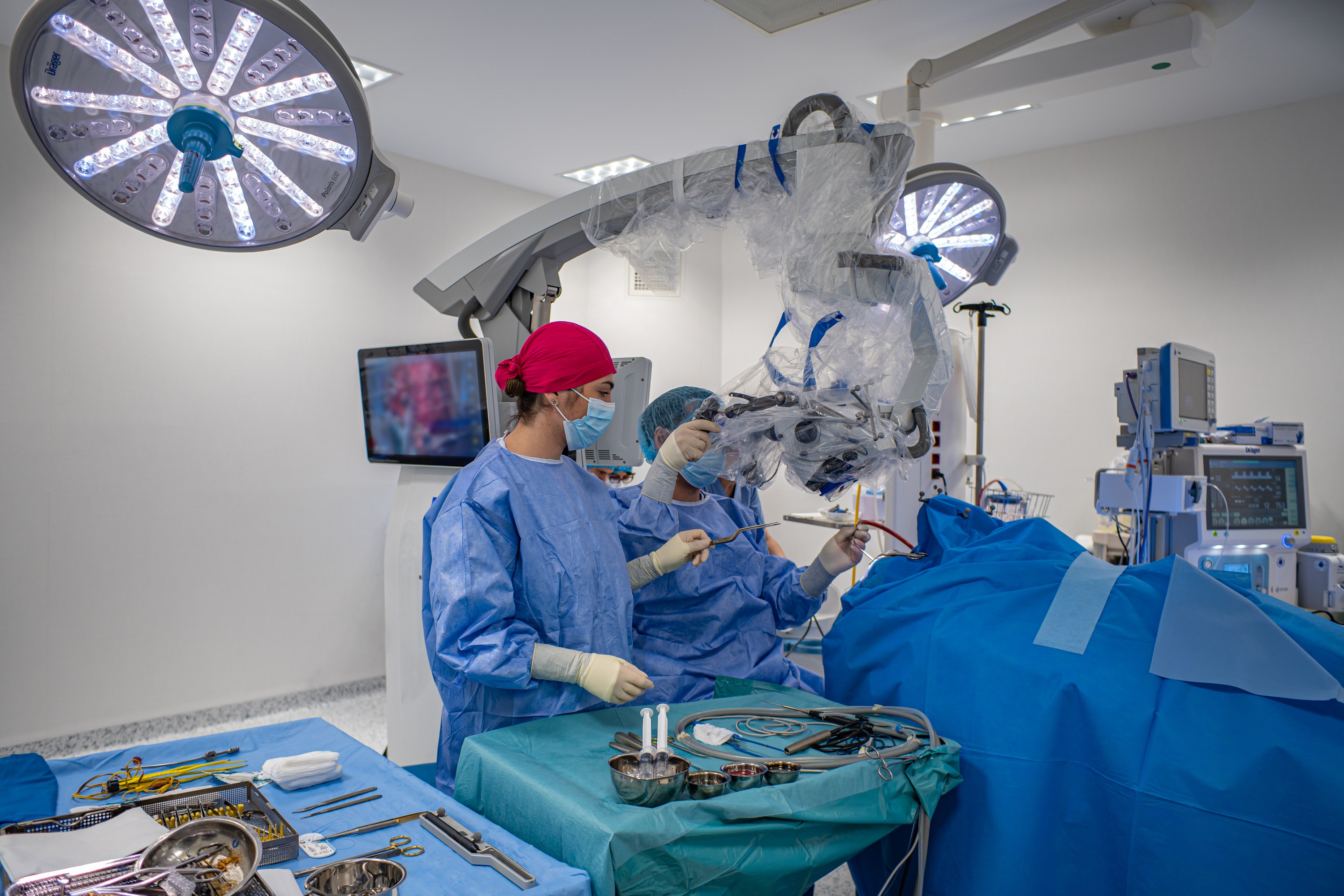
Medical technology helps surgeons achieve efficiency that is difficult to compare with traditional interventions. Dr. Dorin Bica, a primary care neurosurgery physician at Băneasa Memorial Hospital, compares the current technology to GPS because it helps surgeons to achieve higher quality of medical action, make smaller incisions and recover patients faster. The doctor also presented us with the case of a 67-year-old patient with a brain tumor measuring 4 cm, who was discharged only three days after a minimally invasive intervention.
“The peculiarity of Ms. Sabina M.’s case is related to the location of the tumor in the brain. Diagnosis: fronto-parietal parasagittal meningioma on the right. A benign tumor that somehow compresses the sensory and motor area in the part of the brain where we move and feel the arm and leg, especially the leg. This tumor was invading an important vein that drains all blood from the brain, and its removal would become extremely difficult as it progressed. We were interested in removing the entire tumor to minimize the risk of recurrence. A complex and delicate thing, given that it penetrates a vessel with a blood flow of more than a liter per minute. We managed to do this with the help of modern technologies – 3D MRI, neuronavigation, neuroelectrophysiology and special anesthesia,” says Dr. Doreen Bika, doctor of neurosurgery.
The patient appeared for a neurological examination after sporadic numbness of the leg. After an MRI of the brain, he was diagnosed with a meningioma, a 4 cm benign tumor growing from the meninges. The only therapeutic option for this condition is surgery to remove the tumor. The team of doctors from the Memorial Hospital that moved into the work consisted of Dr. Dorin Bike, a primary care neurosurgery doctor, Dr. Iryna Parashiv-Orban, a neurology specialist, and Dr. Alina Voyka, a specialist in anesthesiology and intensive care.

A second MRI followed, this time in 3D, which allows reconstruction of the head in its entirety and helps doctors prepare a pre-operative plan.
“Any operative intervention requires operative planning. This is done at the planning station, that is, we load the MRI, put the head in the optimal position and look at the relationship with the arteries, veins, with the structures of the brain, with everything. When we know the anatomy of the tumor and the corresponding area, we also go to the operating room. I do this all the time, and I’ve noticed that it works best for me if I repeat this entire anatomy the night before the procedure so that it stays in my memory throughout the night,” adds Dr. Bika.
The team of doctors decided that the patient could undergo a minimally invasive procedure, which involves a smaller incision, faster recovery and does not require shaving of hair – an extremely important element for a good postoperative mental state of the patient. In this endeavor, they relied on the support of the latest generation technologies.
“A surgeon relies on the entire team, from the anesthesiologist, surgical and anesthetist assistants, as well as technology. We use technologies like Waze in surgery. It helps us get to our destination faster and better. In “Memorial” there is also a neurologist who deals with neuroelectrophysiology in the office as standard. We monitor, in fact, the cerebral motor pathways to be as sure as possible that we are protecting the patient’s motor function, and in our patient we also used direct stimulation. We put an electrode on the brain that we stimulate to see where the tumor is in relation to the area where the leg moves, especially. And it gives you additional information to help you in your analysis. So, electrophysiology represents for us the standard for almost any intervention we make in the brain. The second thing or the second standard object or technology that we definitely use is navigation. Without it, we can’t make these small cuts, meaning you can’t be precise enough to know to within a centimeter where the tumor is in the brain. And anesthesia, of course, is important. Deep sedation was performed here, which allows us, when we stimulate, to have a muscle reaction,” explains the neurosurgeon.
The intervention lasted four hours, and doctors managed to completely remove the tumor through an incision of only six centimeters. 5 months after the operation, the patient’s condition is very good, the recovery is almost complete.
How is recovery after brain surgery?
Experts recommend that patients resume work a month after the intervention, even if they feel better earlier. During this period of rest, I can take a long walk, go to the theater or the cinema, but without excesses. In addition, the scar must be protected and cared for to prevent infections. Sports can be resumed after three months, when the wound is closed.
Even if the risk of recurrence in benign brain tumors is less than 5%, patients should undergo MRI every three months for the first two years after the intervention. Then the frequency is reduced to once a year or even every two years.
The first symptoms that indicate such a pathology are headaches, epileptic seizures, difficulties with vision, mobility or speech.
Article supported by Memorial Hospital
Source: Hot News
Ashley Bailey is a talented author and journalist known for her writing on trending topics. Currently working at 247 news reel, she brings readers fresh perspectives on current issues. With her well-researched and thought-provoking articles, she captures the zeitgeist and stays ahead of the latest trends. Ashley’s writing is a must-read for anyone interested in staying up-to-date with the latest developments.